Here you see the image of ice crystals from natural snow that are shaped like needles.
In certain arthritis, when fluid from the joint space is examined under
polarized light microscopy, similar needle shaped crystals can be viewed.
 |
Gout and CPPD(Calcium pyrophosphate dihydrate crystal deposition disease) are two of the major causes of acute monoarthritis (acute inflammation of one joint).
CPPD is also termed as pseudogout due to certain similarities with gout seen during acute presentation. It is necessary to differentate between gout and pseudogout as their presentation varies.
Read further to learn their similarities and differences
Similarities of gout and pseudogout:
- causes of acute monoarthritis (acute inflammation of one joint)
- characterized by intra-articular crystal deposition
- the crystal deposits can be observed by visualizing the joint fluid space under polarized light microscopy
- acute episodes of intense pain, redness and visible swelling of the joints involved
- acute episodes triggered by illness, trauma or surgery
- systemic symptoms such as fever maybe present but is not usual.
Differences between gout and pseudogout:
|
Cause of arthritis
|
GOUT
|
PSEUDOGOUT/CPPD
|
|
Crystals
composed of
|
Uric acid/monosodium urate/MSU
|
Calcium pyrophosphate dihydrate/CPPD
|
|
Most
common joint involved
|
1st metacarpophalangeal joint
|
Knee joint
|
|
Polarized
light microscopy
|
Crystals are often intra-cellular, needle-shaped and
yellow
(negatively birefringent)
|
Crystals are rhomboid shaped and blue
(weakly positively birefringent)
|
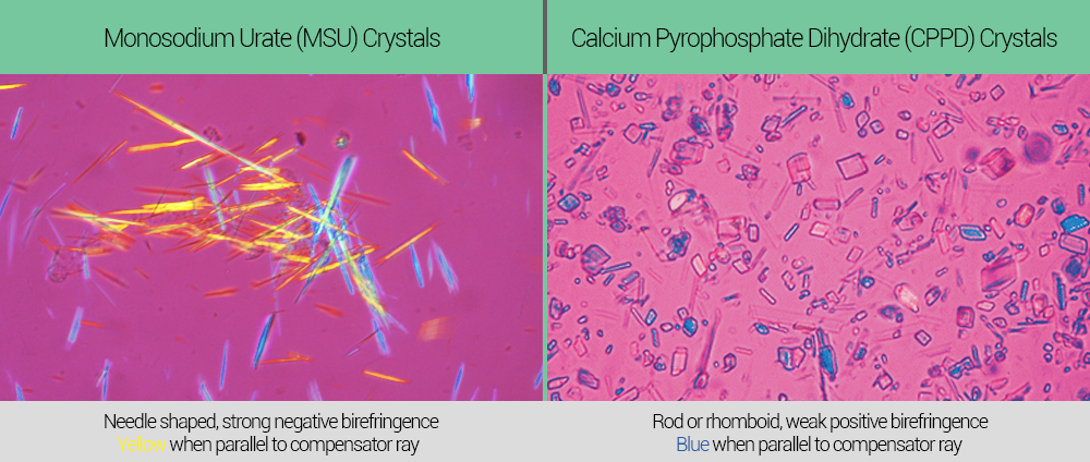
- Gout and pseudogout/CPPD can be differentiated and a definitive diagnosis can be made using polarized light microscopy.
- The image below depicts the difference between the microscopic images of gout and pseudogout/CPPD.

source:www.wikidoc.org
- Note the needle shaped, yellow crystals of uric acid/MSU in gout and rhomboid shaped, blue crystals of CPPD in pseudogout.
Click below links for useful references:
Differences
Orthopaedic notes
Comments
Post a Comment